For decades, researchers have been trying to develop vaccines against cancer. The basic idea sounds almost too simple: teach the immune system what to attack and let it take care of the rest. In practice, however, cancer has turned out to be a much harder target than a virus or a bacterium.
One reason is that cancer is not one disease. Different cancer types have very different molecular characteristics, but even tumours of the same type can vary considerably from one patient to the next. On top of that, these differences are not fixed, because tumours keep evolving as they acquire new mutations and adapt to pressure from treatment and the immune system (McGranahan et al., 2016).
That makes cancer vaccination difficult, but the same diversity also creates an opportunity. If every tumour carries its own molecular fingerprint, we might be able to use that fingerprint to find targets that are specific to one patient’s cancer.
This is the idea behind personalised cancer vaccines, an approach that is now moving quickly towards the clinic. In August 2026, Merck and Moderna announced that their individualised mRNA therapy intismeran autogene, given together with the immunotherapy pembrolizumab, reduced recurrence compared with pembrolizumab alone in a phase 3 trial of more than 1,100 patients whose high-risk melanoma had been surgically removed (NCT05933577). The full results still have to be presented, but according to the companies it is the first positive phase 3 result for this type of therapy. After decades of research on therapeutic cancer vaccines, the field is clearly shifting towards approaches in which the patient’s own tumour is used to design the treatment.
From a Mutation to a Target
One of the most promising strategies is to target neoantigens, which are altered protein fragments that arise from mutations in the tumour and that T cells can potentially recognise. In principle the idea is quite straightforward: we take a sample from the patient’s tumour, sequence it to find the mutations that are unique to the cancer and use that information to design a vaccine containing a selection of targets (Figure 1).

There is an important biological step in between, though, because a mutation does not automatically become a neoantigen. To see why, it helps to follow what happens inside a tumour cell.
As a tumour develops, its cells pick up mutations, some of which fall in genes that encode proteins and change the sequence of amino acids in those proteins. Cells constantly break their proteins down into small fragments called peptides, and some of these peptides are loaded onto HLA molecules that carry them to the cell surface. You can think of HLA molecules as a kind of display system that shows small pieces of the cell’s proteins on the outside, so that passing T cells can inspect them (Figure 2). It is the immune system’s way of getting a glimpse of what is going on inside a cell.
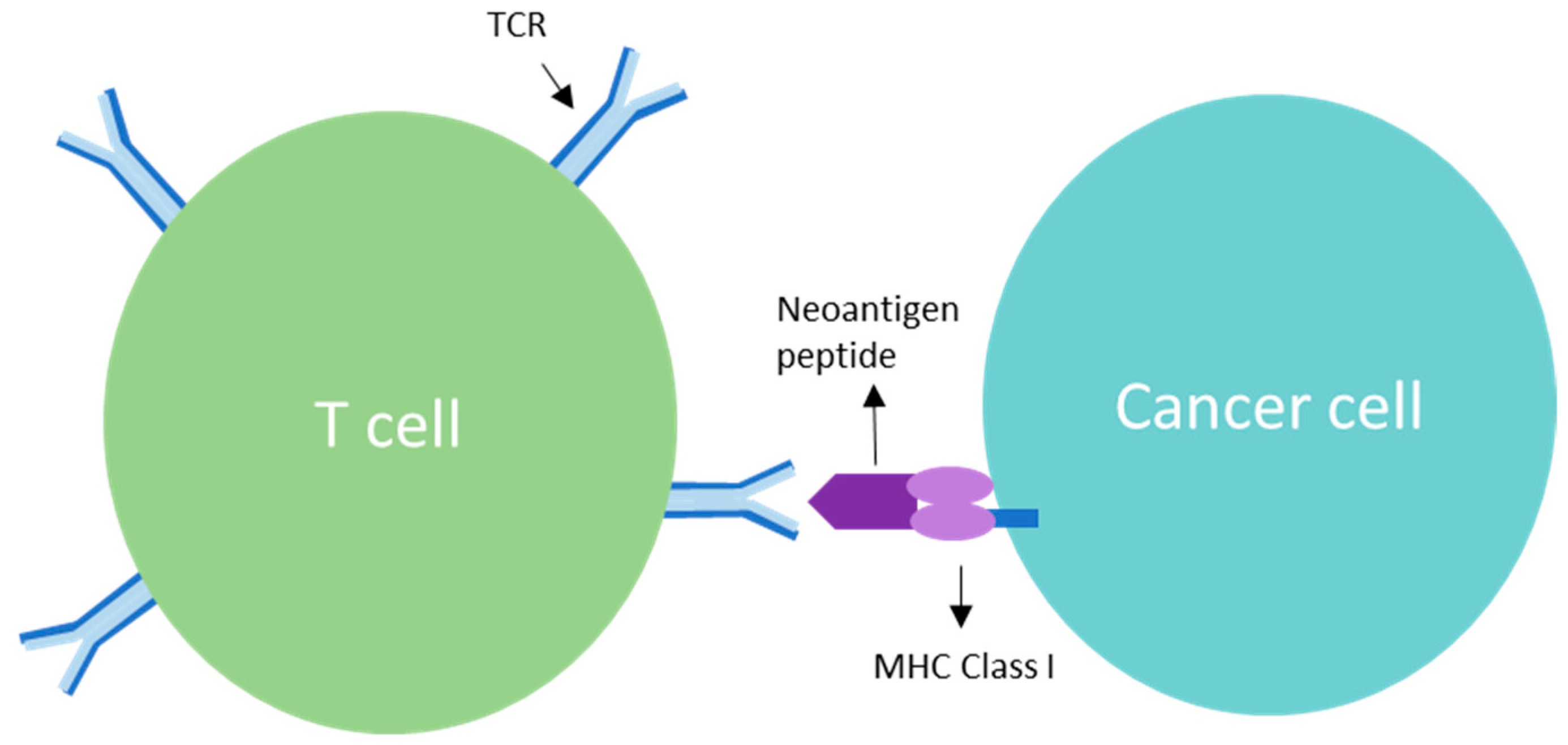
When the displayed peptide comes from a normal protein, the immune system generally recognises it as part of the body. If a mutation has changed the peptide, however, it may look different enough for a T cell to see it as foreign, and that is what we call a neoantigen.
The key word in all of this is potentially. Not every mutation changes a protein and not every altered protein produces a peptide that ends up on HLA. Even when a peptide is displayed, there may be no T cell that recognises it. Working out which mutations will actually make it through all of these steps is where the computational challenge begins.
A Vaccine for One Patient: The Study
In February 2026, a team from BioNTech led by Uğur Şahin and Özlem Türeci published a small but unusually detailed study in Nature (Sahin, Schmidt et al., 2026). They treated 14 patients with triple-negative breast cancer using a vaccine designed from each patient’s own tumour and then followed their immune responses for up to six years.
Triple-negative breast cancer lacks the three receptors that most breast cancer drugs target, which leaves fewer treatment options. Even when it is caught early, it is more likely to come back than other types of breast cancer (Li et al., 2017). All patients in the study had already completed their standard treatment and had no visible cancer left, so the vaccine was not meant to shrink a tumour but to teach the immune system to recognise any cancer cells that might still be hiding.
Choosing the targets
For each patient, the team sequenced the tumour and compared it with DNA from healthy cells to find the mutations that were unique to the cancer. Prediction algorithms, including tools from the Immune Epitope Database, then estimated which of these mutations were most likely to be displayed on the patient’s own HLA molecules. Because HLA genes differ a lot between people, this step is personal as well: a mutation that is visible to the immune system in one patient may be invisible in another. Based on this ranking, up to 20 mutations were selected per patient, after which a team of experts checked every list by hand.
From targets to vaccine
The selected mutations were then written into mRNA, which is essentially a set of instructions telling cells to produce small pieces of the mutated proteins. The mRNA is packaged in tiny fat particles and given through an infusion, after which it ends up mainly in the lymph nodes and spleen where T cells are trained. BioNTech had already tested the same platform in melanoma (Sahin et al., 2017) and pancreatic cancer (Rojas et al., 2023). Producing each personal vaccine took a little over two months, after which patients received eight doses over roughly nine weeks.
But doesn’t the tumour already carry these mutations?
It does, which raises the question of why the immune system needs a vaccine at all. The answer is that seeing a target is not the same as learning to attack it. T cells are trained in the lymph nodes by specialised cells, but only when there are clear signs of danger, which a tumour rarely provides. The vaccine delivers the same targets to the right place in the right way, so that T cells can learn to recognise them before going out to find the tumour cells that carry them.
This is also what the study found: most of the T-cell responses were new and could not be detected before vaccination, even though the patients had been carrying these mutations all along.
What Did They Find?
The first and perhaps most striking result is that every patient responded. All 14 patients developed T cells against at least one of their vaccine targets and 13 of them against several.
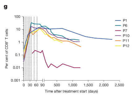
The responses were also strong. In some patients more than one in ten of their killer T cells recognised a single vaccine target after vaccination, which the authors point out is a level usually only seen with T-cell therapies in which cells are grown in the lab and infused back into the patient.
Finally, the responses lasted. Even without booster shots, vaccine-induced T cells remained detectable in the blood for years (Figure 3). In one patient they were still present six years later, including cells that can renew themselves and rebuild the response when needed.
Three Relapses, Three Lessons
Eleven of the 14 patients remained free of relapse for up to six years (Figure 4). The three patients who did relapse are perhaps the most interesting part of the paper, because each of them shows a different way in which this approach can fail.
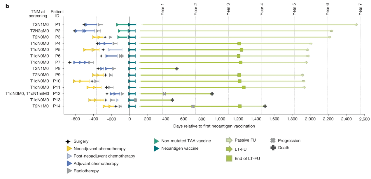
In the first patient, the vaccine simply didn’t take. She had a very weak immune response and her cancer returned, but she then responded to a different type of immunotherapy, with the cancer disappearing for more than a year. That suggests that combining the two approaches could help patients whose immune system responds poorly to the vaccine alone.
The second patient had a strong response, with vaccine-induced T cells that even reached the returning tumour. The tumour, however, had stopped displaying its peptides on HLA, which meant that the T cells could no longer recognise it.
In the third patient, the relapse came from a different tumour altogether. She had tumours in both breasts but only one of them was used to design the vaccine. The relapse turned out to come from the other one, which carried a completely different set of mutations, a reminder that every tumour is different even within a single patient.
A Word of Caution
It is important to keep in mind that this is an early-stage study. With only 14 patients and no comparison group, it cannot tell us whether the vaccine actually prevented relapses. Patients whose cancer returned before their vaccine was ready could also not take part, which may make the results look better than they are. Answering that question will require larger randomised trials such as the Merck and Moderna study in melanoma.
The computational challenge is also far from solved. Of all the mutations included in the vaccines, only about one in six triggered an immune response strong enough to measure directly in the blood. Prediction gives us a useful shortlist, but many of the predicted targets turn out to stay silent.
Towards Personalised Oncology: From Vaccines to Digital Twins
This study fits into a broader shift in computational oncology from asking what works on average to asking what will work for a specific patient. Here, sequencing and prediction together determine what goes into each vaccine, yet the relapses show that predicting targets is not the same as predicting outcomes. Understanding why one patient barely responded or why a tumour stopped displaying its mutations requires knowing how a tumour and an immune system interact over time, which a single snapshot of a tumour cannot capture.
This is where digital twins come in. A digital twin is a computational model of an individual patient’s disease that is built from that patient’s own data and updated as new data comes in, so that it can be used to simulate how the disease might respond to different treatments before they are given. The concept comes from engineering, where digital twins of aircraft engines are used to anticipate failures before they happen. In oncology, the US National Cancer Institute and Department of Energy launched a joint initiative on cancer patient digital twins in 2020 (Stahlberg et al., 2022) and researchers have since proposed frameworks in which such twins keep learning from clinical data to support treatment decisions (Hernandez-Boussard et al., 2021). Applied to this study, a digital twin could combine a patient’s tumour sequencing, immune profile and clinical data to explore scenarios like the three relapses, for example a small group of tumour cells that stops displaying its mutations, and to test whether adding another therapy would change the outcome.
Such models are still a long way from the clinic, as they require high-quality data, careful validation and an honest estimate of their own uncertainty. The direction is clear, though: as treatments become more personal, the models that guide them will have to become more personal too.
The Bottom Line
This study does not prove that personalised mRNA vaccines prevent breast cancer from coming back. What it does show is that a vaccine can be designed for a single patient’s tumour within a clinical timeline, that it consistently teaches the immune system to recognise that tumour and that this recognition can last for years without boosters. The failures turn out to be just as informative as the successes, because they show that personalisation will have to go beyond a single snapshot of a single tumour towards understanding how each patient’s disease changes over time.
Every tumour is different, which is exactly why the way we treat it is becoming different too.
Read More
- Original study (open access): Sahin, Schmidt et al. (2026), Nature
- Clinical trial: TNBC-MERIT, NCT02316457
- Phase 3 melanoma trial announcement: Merck and Moderna, August 2026
- Cancer patient digital twins: Hernandez-Boussard et al. (2021), Nature Medicine
Related research project within SEEDBiomed network
B. Brouwer, V. M. Sheraton and A. Hoekstra (University of Amsterdam), together with M. Haselager and E. Eldering (Amsterdam UMC), are building a digital twin of patient-derived 3D spheroids for chronic lymphocytic leukaemia (CLL) and related B-cell lymphomas. In this project, cancer cells from patients are grown as small 3D clusters with and without T cells, imaged over time and exposed to drugs. AI-based image analysis and RNA sequencing turn these experiments into quantitative features that are used to calibrate a multiscale simulation of the spheroid, with the aim of predicting how an individual patient’s cancer will respond to treatment and, eventually, why it might stop responding.